


Table of Contents
- QUICK SUMMARY
- Introduction
- What Is Ocular Melanoma? (Eye Cancer Melanoma Explained)
- What Are the Early Ocular Melanoma Symptoms to Watch Out For?
- What Causes Ocular Melanoma? Risk Factors Explained
- How Is Ocular Melanoma Diagnosed? Tests and Procedures Explained
- Uveal Melanoma Treatment Options in 2026
- Ocular Melanoma Treatment Cost in India (2026 Guide)
- Can’t Afford Ocular Melanoma Treatment? Here’s How Families Are Getting Financial Help
- Conclusion
- FAQs
QUICK SUMMARY
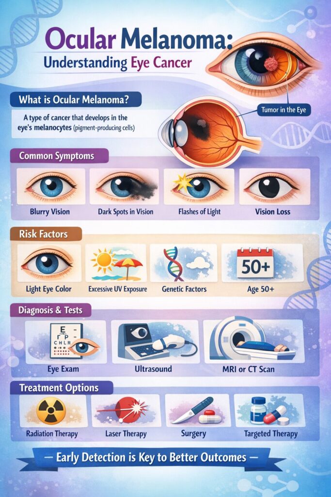
- Ocular melanoma is the most common primary eye cancer in adults, developing in the pigment-producing cells (melanocytes) inside or around the eye.
- Early symptoms such as blurred vision, floaters, a dark spot on the iris, or changes in pupil shape are often absent in the initial stages, making routine eye exams critical.
- Treatment options include plaque brachytherapy, proton beam therapy, and surgery; early-stage ocular melanoma has a significantly better prognosis.
- Ocular melanoma treatment in India costs between ₹2 lakh and ₹8.3 lakh.
- Families managing the financial burden of eye cancer treatment in India can explore medical crowdfunding as a practical, zero-debt option for raising funds.
Introduction
A cancer diagnosis is always life-changing. When that diagnosis is ocular melanoma, a cancer that silently grows inside the eye, often without any warning signs, it can feel especially overwhelming.
Ocular melanoma, also called eye cancer melanoma, is the most common primary eye cancer in adults. While it is rare globally, it is serious: without early detection and timely treatment, it can spread to the liver, lungs, and other organs, significantly reducing survival odds.
In India, ocular melanoma is diagnosed less frequently than in Western populations. Still, the cases that do occur are often detected late, partly because awareness remains low and partly because early symptoms are easy to dismiss. That is what this guide aims to change. It covers everything, from what ocular melanoma is and how it develops, to what diagnosis and treatment look like in India today, and how families can get financial help for medical treatment when costs reach out of pocket.
What Is Ocular Melanoma? (Eye Cancer Melanoma Explained)
Quick Answer: Ocular melanoma is a rare cancer that develops from melanocytes, the pigment-producing cells, in or around the eye. It is the most common primary eye cancer in adults and the second most common form of melanoma after skin melanoma. It is one of the rarest cancers in India
Melanoma is usually known as a skin cancer, but the same pigment-producing cells that give skin its colour, called melanocytes, are also present in the eye. When these cells mutate and begin to grow uncontrollably, ocular melanoma forms.
Most cases develop in the uvea, the middle layer of the eye that lies between the outer white sclera and the inner retina. Because this part of the eye is not visible from the outside, tumours can grow for months or even years without being noticed, not by the patient, and sometimes not even by a general physician.
According to data from the International Agency for Research on Cancer (IARC) and the SEER database, the global incidence of ocular melanoma is estimated between 4.3 and 5.1 cases per million people annually. The incidence in India is significantly lower than in Western nations. Clinical research indicates that age-adjusted rates in India fall below 1.0 case per million person-years, whereas rates exceed 6.0 in Northern Europe, the USA, Canada, and Australia.
Types of Ocular Melanoma
Ocular melanoma is not a single disease; it is an umbrella term for melanoma that develops in different parts of the eye. Understanding the type matters because it directly affects symptoms, treatment approach, and prognosis.
1. Uveal Melanoma
Uveal melanoma is the most common type of ocular melanoma, developing from melanocytes within the uveal tract, which includes the iris (the coloured part of the eye), the ciliary body (the ring of muscle that controls the lens), and the choroid (the layer of blood vessels lining the back of the eye).
Within uveal melanoma, there are three subtypes based on location:
- Choroidal Melanoma – The most common subtype. The majority of uveal melanomas originate from the choroid. Because the choroid sits at the back of the eye, tumours here are the hardest to detect early.
- Ciliary Body Melanoma – Arises in the ring of muscle around the lens. It accounts for roughly 10% of uveal melanomas and tends to be more aggressive.
- Iris Melanoma – The rarest subtype, and the one most visible to patients (and doctors), as it appears as a dark spot on the coloured part of the eye. It generally carries a better prognosis than choroidal or ciliary body melanoma.
2. Conjunctival Melanoma
Less common than uveal melanoma, conjunctival melanoma develops on the conjunctiva, the thin, transparent membrane that covers the white of the eye and lines the inside of the eyelids. Unlike uveal melanoma, conjunctival melanoma shares genetic similarities with skin melanoma, often featuring TERT promoter, NRAS, and NF1 mutations. It is rarer, but its surface location means it is sometimes noticed earlier.
Ocular Melanoma vs Uveal Melanoma: What Is the Difference?
This is one of the most commonly searched questions around this topic, so let’s address it directly.
- Ocular melanoma is the broader term; it refers to any melanoma that occurs in or around the eye, including both uveal and conjunctival melanoma.
- Uveal melanoma is a specific subtype of ocular melanoma, and the most common one. So while all uveal melanomas are ocular melanomas, not all ocular melanomas are uveal melanomas.
When most doctors, researchers, or articles refer to “ocular melanoma,” they are typically referring to uveal melanoma, which is why the two terms are often used interchangeably, even though they are technically distinct.
What Are the Early Ocular Melanoma Symptoms to Watch Out For?
Quick Answer: Ocular melanoma is often called a “silent cancer” because it may cause no symptoms in its early stages. When symptoms do appear, they typically include blurred vision, floaters, flashes of light, or a growing dark spot on the iris. Any sudden or unexplained change in vision should be evaluated by an ophthalmologist without delay.
One of the most challenging aspects of ocular melanoma is how quietly it begins. Early-stage ocular melanoma often causes no symptoms and may only be detected during a routine eye exam. This is especially true for tumours developing in the choroid, the back layer of the eye, where growth can go completely undetected until the tumour reaches a size that begins to affect vision.
This is why eye specialists and oncologists consistently emphasise one thing above all else: regular, comprehensive eye examinations with pupil dilation are the single most important tool for early detection.
Common Ocular Melanoma Symptoms – A Detailed Breakdown
As a tumour grows larger, it begins to press on or displace surrounding eye structures, and symptoms start to emerge.
Here is what each symptom looks like in practice:
- Blurred or Distorted Vision: Vision may become hazy, wavy, or distorted, particularly in one eye. This happens when the tumour grows large enough to affect the retina or the fluid dynamics inside the eye.
- Floaters and Flashes of Light: Spots or squiggles drifting in the field of vision, or flashes of light, are among the recognised warning signs of ocular melanoma. While floaters are extremely common and usually harmless (especially with ageing), a sudden increase in their number or the appearance of new floaters alongside flashes of light warrants prompt evaluation.
- A Growing Dark Spot on the Iris or Sclera: Dark spots on either the white of the eye (sclera) or the iris are among the more common symptoms of ocular melanoma. Any mole or dark patch on the eye that appears new or is visibly growing should be assessed immediately.
- Change in Pupil Shape or Size: A change in the shape or size of the pupil, the dark circle in the middle of the iris, is a recognised symptom of ocular melanoma.
- Loss of Peripheral Vision: As the tumour enlarges, it can encroach on the retina or optic nerve pathways, causing blind spots or a narrowing of the visual field, often in one eye only.
- Eye Pain or Pressure: In advanced cases, rising intraocular pressure (a condition called secondary glaucoma, caused by the tumour blocking normal fluid drainage) can result in a persistent aching or pressure sensation in the eye.
- Changes in Eye Position or Movement: Changes in the eyeball’s position or movements within the socket. It can occur in more advanced or larger tumours, where the mass begins to physically shift the eye’s placement.
Ocular Melanoma Symptoms by Tumour Location
Symptoms also vary depending on where exactly inside the eye the melanoma has developed:
| Location | Likely Symptoms |
| Choroid (back of eye) | Blurred vision, floaters, and retinal detachment are often asymptomatic early |
| Ciliary Body | Lens displacement, blurred vision, secondary glaucoma |
| Iris | Visible dark spot, change in pupil shape, most easily noticed |
| Conjunctiva | Visible pigmented lesion on the eye surface, redness, irritation |
When Should You See a Doctor?
Any of the following should prompt an urgent visit to an ophthalmologist; do not wait for a scheduled appointment:
- A sudden increase in floaters or new flashes of light
- Unexplained blurred or distorted vision in one eye
- A new or growing dark spot on the iris or the white of the eye
- A noticeable change in the shape of your pupil
- Gradual loss of side vision in one eye
It is also worth repeating: some eye cancers are detected during routine eye exams without the patient having experienced any symptoms at all, making regular, dilated eye exams non-negotiable, particularly for anyone over the age of 40 or with known risk factors.
What Causes Ocular Melanoma? Risk Factors Explained
Quick Answer: The exact cause of ocular melanoma is not fully understood, but it begins when DNA mutations occur in the melanocytes (pigment cells) of the eye, causing them to multiply uncontrollably. Key risk factors include light-coloured eyes, fair skin, UV radiation exposure, and specific genetic mutations, particularly in the BAP1 gene.
1. UV Radiation and Sunlight Exposure
Exposure to ultraviolet (UV) radiation is a recognised risk factor for ocular melanoma, although the link is less direct than with skin melanoma. People with high occupational exposure, such as welders, may have a higher risk.
2. Light Eye Colour and Fair Skin
Individuals with light-coloured eyes (blue, green, or grey) and fair skin have a higher risk of eye cancer melanoma. Lower melanin levels in the eye may reduce natural protection against UV-related damage. This is one of the primary reasons why ocular melanoma is significantly more common in Caucasian populations than in people of Asian or African descent.
3. The BAP1 Gene Mutation
The BAP1 gene mutation is the most significant known genetic risk factor for ocular melanoma. This mutation affects the body’s ability to control abnormal cell growth and is linked to more aggressive forms of the disease.
In rare cases, it occurs as part of an inherited condition (BAP1 tumour predisposition syndrome), which may increase the risk of multiple cancers. Genetic counselling is advised for individuals with a strong family history.
4. Pre-existing Ocular Moles (Nevi)
Certain pigmented lesions in the eye (nevi), including choroidal or iris nevi, may increase the risk of developing uveal melanoma. These should be regularly monitored during eye examinations.
5. Age
Ocular melanoma is more common in older adults, with risk increasing significantly after age 60. However, it can occur at any age in rare cases.
6. Dysplastic Nevus Syndrome
Individuals with certain inherited conditions, such as dysplastic nevus syndrome, face an increased risk of ocular melanoma. This condition, characterised by the presence of unusual moles on the skin, has been associated with a higher likelihood of developing both skin and eye melanoma.
7. Primary Acquired Melanosis (PAM)
Primary acquired melanosis (PAM) is a condition involving abnormal pigmentation of the conjunctiva and is a known risk factor for conjunctival melanoma. This should be monitored closely by an ophthalmologist.
Is Ocular Melanoma Hereditary?
This is a question many patients and families ask after diagnosis, and the answer is: mostly no, but sometimes yes.
- Familial uveal melanoma, defined as two or more family members diagnosed with the condition, is rare and estimated at less than 1% of all uveal melanoma cases. Currently, BAP1 is the only gene known to contribute significant risk of uveal melanoma in a hereditary pattern.
- The vast majority of ocular melanoma cases are sporadic, meaning they arise from random mutations with no family connection.
- However, if you have a first-degree relative (parent, sibling, or child) who has been diagnosed with uveal melanoma, mesothelioma, renal cell carcinoma, or cutaneous melanoma, it is worth discussing BAP1 genetic testing with your doctor.
Early knowledge of a BAP1 mutation can significantly change surveillance and prevention strategies for the whole family.
Can Ocular Melanoma Be Prevented?
There is no guaranteed way to prevent ocular melanoma, particularly since the most significant risk factors (eye colour, skin tone, genetics) are outside our control. However, there are sensible steps worth taking:
- Wear UV-protective sunglasses outdoors, especially wraparound styles that block UVA and UVB rays
- If you work as a welder or in environments with intense UV exposure, use appropriate protective eyewear
- Schedule regular, dilated eye exams annually if you are over 60, or more frequently if you have known risk factors
- If you have a family history of BAP1-associated cancers, speak to a genetic counsellor
How Is Ocular Melanoma Diagnosed? Tests and Procedures Explained
Quick Answer: Ocular melanoma is primarily diagnosed through a clinical eye examination, not a biopsy. A dilated fundus exam, ocular ultrasound, and imaging tests like MRI or CT scan are the standard diagnostic tools. Genetic testing of a tumour biopsy is increasingly used to assess metastatic risk rather than to confirm the diagnosis itself.
Clinical Eye Examination
The diagnostic process typically begins with a comprehensive dilated eye exam performed by an ophthalmologist.
During this exam, doctors look for:
- Abnormal pigmented lesions inside the eye
- Changes in retinal structure
- Tumour size, shape, and location
Many cases of ocular melanoma diagnosis happen during routine eye check-ups, even before symptoms appear.
Imaging Tests Used in Diagnosis
Imaging plays a central role in confirming eye cancer, melanoma, and understanding its extent.
- Ocular Ultrasound (A-scan and B-scan): Helps determine tumour size, thickness, and internal characteristics
- Optical Coherence Tomography (OCT): Provides high-resolution images of retinal layers and detects fluid buildup
- MRI or CT Scan: Used to evaluate whether the cancer has spread beyond the eye, especially to the liver or lungs
These tests together help confirm ocular melanoma diagnosis and guide treatment planning.
Is Biopsy Required?
- Not always needed for initial diagnosis
- May be performed in selected cases
- Primarily used for genetic and molecular testing
Biopsy helps determine:
- Risk of metastasis
- Tumour aggressiveness
- Suitability for targeted therapies
Role of Genetic Testing
Genetic testing is increasingly used in uveal melanoma diagnosis to assess future risk.
Key insights include:
- Likelihood of cancer spreading
- Need for closer follow-up or systemic treatment
Why Early Diagnosis Matters
Early detection significantly improves outcomes in ocular melanoma treatment.
- Higher chances of preserving vision
- Lower risk of metastasis
- More treatment options available
Regular eye exams remain the most effective way to detect ocular melanoma early.
Ocular Melanoma Stages
Ocular melanoma is staged according to the AJCC 8th Edition staging system, which considers tumour thickness, basal diameter, ciliary body involvement, and whether the tumour has extended beyond the eye.
| Stage | What It Means |
| Stage I (T1) | Small tumour, confined to the eye, low metastatic risk |
| Stage II (T2) | Medium tumour, confined to the eye |
| Stage III (T3–T4) | A larger tumour may involve the ciliary body or extend beyond the eye |
| Stage IV | Distant metastasis — most commonly to the liver |
AJCC staging is no longer sufficient by itself for modern prognostication; molecular and cytogenetic data substantially refine risk prediction and should be combined with anatomical staging for a complete picture.
Uveal Melanoma Treatment Options in 2026
Quick Answer: Ocular melanoma treatment depends on tumour size, location, and spread. For most small-to-medium tumours, eye-preserving radiation therapy is the first-line approach. Surgery is used for larger tumours, while systemic therapies are considered in advanced cases.
The goal of ocular melanoma treatment is to remove or control the tumour while preserving vision whenever possible.
Radiation Therapy (First-Line Treatment)
Radiation therapy is the most common treatment for uveal melanoma, especially in early and mid-stage disease.
- Plaque brachytherapy: A targeted form of radiation placed near the tumour for a few days to destroy cancer cells while preserving the eye
- Proton beam therapy: A highly precise radiation option used for larger tumours or those near critical eye structures
Both treatments offer comparable survival outcomes and are widely used in modern ocular melanoma treatment.
Surgical Treatment
Surgery is considered when radiation is not suitable or the tumour is advanced.
- Local resection: Removal of the tumour while preserving the eye in selected cases
- Enucleation: Removal of the entire eye, typically for large tumours or when vision cannot be preserved
Treatment for Metastatic Disease
If eye cancer melanoma spreads (most commonly to the liver), systemic treatment is required.
- Tebentafusp: The only approved targeted therapy for metastatic uveal melanoma, shown to improve survival in eligible patients
- Liver-directed therapies: Used when cancer is confined to the liver
- Immunotherapy and clinical trials: Emerging options being studied to improve outcomes in advanced disease

Ocular Melanoma Treatment Cost in India (2026 Guide)
Quick Answer: Ocular melanoma treatment in India costs between ₹2 lakh and ₹8.3 lakh on average, compared to ₹25 lakh to ₹60 lakh in the USA. The exact cost depends on treatment type, hospital, city, and tumour stage. India offers internationally accredited care at a fraction of Western prices, making it an increasingly sought-after destination for eye cancer treatment.
India has made significant strides in ocular oncology, including the development of a cost-effective make-in-India Ruthenium-106 plaque for treating ocular tumours via surface plaque brachytherapy, a home-grown innovation that has helped reduce treatment costs significantly for Indian patients.
Cost Breakdown by Treatment Type in India (2026)
| Treatment | Estimated Cost (INR) | Estimated Cost (USD) |
| Plaque Brachytherapy | ₹2,00,000 – ₹5,00,000 | $2,400 – $6,000 |
| External Beam Radiation Therapy | ₹1,50,000 – ₹3,50,000 | $1,800 – $4,200 |
| Proton Beam Therapy | ₹8,00,000 – ₹15,00,000 | $9,600 – $18,000 |
| Local Resection Surgery | ₹2,50,000 – ₹5,00,000 | $3,000 – $6,000 |
| Enucleation (Eye Removal) | ₹80,000 – ₹1,50,000 | $950 – $1,800 |
| Laser Therapy / Cryotherapy | ₹60,000 – ₹1,50,000 | $720 – $1,800 |
| Immunotherapy (per cycle) | ₹1,50,000 – ₹3,00,000 | $1,800 – $3,600 |
Note: Costs above cover the procedure itself. Diagnostic tests, hospital stay, and follow-up imaging add to the total.
What Factors Affect the Cost of Treatment in India?
- What Factors Affect Ocular Melanoma Treatment Cost?
- Type of treatment: Proton therapy is the most expensive; surgery and laser options are lower-cost
- Tumour size and stage: Advanced cases require more complex treatment
- Hospital and city: Costs are higher in metro cities and private hospitals
- Diagnostic tests: MRI, CT scans, and genetic testing add to overall expenses
- Insurance coverage: Varies by policy, especially for advanced therapies
Leading Hospitals for Ocular Melanoma Treatment in India
India has a strong network of specialised eye and cancer centres equipped to diagnose and treat ocular melanoma across all stages, from early detection to advanced care.
- Sankara Nethralaya, Chennai: A leading eye hospital with a dedicated ocular oncology department experienced in treating uveal melanoma, including choroidal melanoma. Offers advanced treatments such as plaque brachytherapy, laser therapy, and local radiotherapy.
- LV Prasad Eye Institute, Hyderabad: A globally recognised comprehensive eye care centre with a specialised ocular oncology programme. Known for high-quality care and accessibility, with a significant portion of patients receiving free or subsidised treatment.
- AIIMS (Rajendra Prasad Centre for Ophthalmic Sciences), New Delhi: One of India’s top government institutions for eye care, offering advanced diagnostic and treatment facilities for eye cancer and melanoma, along with subsidised treatment options for eligible patients.
- Tata Memorial Hospital, Mumbai: A premier cancer treatment centre in India, well-equipped to manage complex and metastatic ocular melanoma cases, including those requiring systemic therapy and multidisciplinary care.
- Apollo Proton Cancer Centre, Chennai: The first proton therapy centre in South Asia, offering proton beam therapy for ocular melanoma, particularly beneficial for tumours located near critical eye structures.
Can’t Afford Ocular Melanoma Treatment? Here’s How Families Are Getting Financial Help
While ocular melanoma treatment in India is more affordable than in many countries, costs ranging from ₹2 lakh to ₹8.3 lakh can still be overwhelming for many families. Expenses such as diagnostics, hospital stays, follow-up scans, and advanced therapies can quickly increase the total financial burden.
With a significant portion of the population lacking comprehensive health insurance, many families are forced to delay treatment or take on debt. To bridge this gap, medical crowdfunding has emerged as a practical option for raising funds quickly and transparently.
Medical crowdfunding allows patients and families to raise donations online by sharing their story with a wider community. Instead of relying on loans, contributions from multiple donors help cover treatment costs without repayment, making it a viable option during medical emergencies.
How ImpactGuru Helps Families Fund Eye Cancer Treatment
ImpactGuru is one of India’s leading online crowdfunding platforms for healthcare, supporting thousands of patients across the country.
- 0% platform fee, allowing more funds to go directly toward treatment
- Ability to start a fundraiser within minutes
- Access to 24/7 support and campaign guidance
- Funds are transferred directly to verified bank accounts for transparency
Families can use crowdfunding to manage expenses related to ocular melanoma treatment, including radiation therapy, surgery, diagnostic tests, and follow-up care.
Conclusion
Ocular melanoma is rare, but it is serious, and its silence in the early stages makes routine eye examinations genuinely life-saving. Whether you have risk factors or not, a dilated eye exam every one to two years remains the single most reliable way to catch this cancer before it spreads.
India today offers world-class ocular melanoma care, but if cost is still a barrier, medical crowdfunding platforms in India like ImpactGuru have helped over 50,000 patients access timely treatment without taking on debt.
Medical Disclaimer: This article is intended for informational purposes only and should not be construed as medical advice. Always consult a qualified ophthalmologist or oncologist for diagnosis and treatment decisions.
FAQs
Ocular melanoma is a rare type of cancer that develops in the pigment-producing cells (melanocytes) of the eye, most commonly in the uvea (middle layer).
Early symptoms may include blurred vision, flashes of light, floaters, or a growing dark spot on the iris. Some patients may have no symptoms initially.
The exact cause is unknown, but it is linked to genetic mutations in eye cells that lead to uncontrolled growth of melanocytes.
People over 50, those with light-colored eyes, fair skin, or certain genetic conditions are at higher risk.
Yes, it is a serious cancer that can spread to other organs, especially the liver, if not treated early.
It is usually diagnosed through a detailed eye examination, ultrasound imaging, and sometimes scans like MRI or CT.
Treatment may include radiation therapy, laser therapy, surgery, or targeted treatments depending on tumor size and location.
You should consult an eye specialist if you notice sudden vision changes, flashes, floaters, or dark spots in your eye.