
Craniosynostosis is a medical condition characterized by the premature fusion of one or more sutures, or joints, between the bones of an infant’s skull. Typically, these sutures remain open during early childhood, allowing the skull to expand and accommodate the growing brain. However, in cases of craniosynostosis, the fusion of these sutures occurs too early, restricting normal skull growth and shaping. This condition can result in an abnormal head shape, potentially leading to complications if left untreated.
The term “craniosynostosis” is derived from two words: “cranio,” meaning skull, and “synostosis,” referring to the fusion of bones. The fusion of the skull bones can affect different areas, giving rise to several types of craniosynostosis. The most common types include sagittal synostosis, which involves the fusion along the top of the skull, and coronal synostosis, where the fusion occurs on one side of the forehead. Other types, such as metopic and lambdoid synostosis, affect different regions of the skull, presenting unique challenges in diagnosis and treatment.
Craniosynostosis is often associated with craniofacial syndromes, genetic disorders affecting the head and face. One such syndrome is Apert syndrome, characterized by the fusion of specific skull bones, leading to distinctive facial and hand abnormalities. Another example is Crouzon syndrome, marked by the early fusion of certain skull bones and a range of facial features.
Understanding the causes of craniosynostosis is crucial for both diagnosis and treatment. While some cases may be idiopathic, with no apparent cause, genetic factors are at play in many instances. Mutations in certain genes have been linked to craniosynostosis; a family history of the condition increases the risk. Additionally, environmental factors, maternal health during pregnancy, and certain medications may contribute to the development of craniosynostosis.
Early detection of craniosynostosis is essential for effective intervention. Diagnosis often involves a combination of physical examination, medical history review, & imaging studies such as CT scans. Timely identification allows for the implementation of appropriate treatment strategies. Surgical intervention is the primary method for managing craniosynostosis, involving the release of fused sutures to allow for normal skull growth. The timing and approach of surgery depend on several factors, including the severity of the condition and the specific type of craniosynostosis.
In conclusion, craniosynostosis is a complex medical condition that affects the normal development of an infant’s skull. With various types and potential syndromic associations, early diagnosis and intervention are crucial to managing the condition effectively. A comprehensive understanding of the causes, types, and treatment options for craniosynostosis is essential for healthcare professionals, parents, and caregivers involved in the care of affected individuals.
Table of Contents
Types Of Craniosynostosis
Craniosynostosis is a medical condition characterized by the premature fusion of one or more of the fibrous joints between the bones of an infant’s skull, which can affect the shape & growth of the head. The medical condition can be classified based on the specific sutures that fuse prematurely. Here are some common types of craniosynostosis:
1. Sagittal Synostosis (Scaphocephaly):
– Location: The sagittal suture runs from the front to the back at the top of the skull.
– Characteristics: Premature fusion of the sagittal suture leads to a long, narrow head shape. The head often appears boat-shaped.
2. Coronal Synostosis (Plagiocephaly):
– Location: The coronal sutures are on the top of the head, running from ear to ear.
– Characteristics: Premature fusion of one or both coronal sutures results in an asymmetric head shape. The forehead and eye sockets on the affected side may appear flattened.
3. Metopic Synostosis (Trigonocephaly):
– Location: The metopic suture is at the top of the forehead, running from the nose to the sagittal suture.
– Characteristics: Premature fusion of the metopic suture causes a triangular or pointed forehead, giving the head a wedge-shaped appearance.
4. Lambdoid Synostosis (Posterior Plagiocephaly):
– Location: The lambdoid sutures are located at the back of the head.
– Characteristics: Premature fusion of the lambdoid sutures can cause flattening on one side of the back of the head.
5. Multiple Suture Synostosis (Complex Craniosynostosis):
– Characteristics: In some cases, more than one suture fuses prematurely, leading to a combination of features from different types of craniosynostosis.
6. Syndromic Craniosynostosis:
– Characteristics: Craniosynostosis can also occur as part of a genetic syndrome, such as Apert syndrome, Crouzon syndrome, Pfeiffer syndrome, or Saethre-Chotzen syndrome. These syndromes often involve abnormalities in other parts of the body, in addition to the skull.
Craniosynostosis Meaning
Craniosynostosis is a medical condition in which one or more joints between the bones of an infant’s skull close prematurely before the brain fully develops. In a normal skull, these joints, known as sutures, allow for gradual skull growth as the brain expands. When one or more sutures close too early, it can lead to problems with normal skull and brain growth.
The premature fusion of the skull bones can result in an abnormal head shape and, in some cases, increased pressure on the brain. This increased pressure can potentially cause developmental issues and affect the shape of the face and head. The cause of craniosynostosis is not always clear, but it can be associated with genetic factors or sporadically.
Treatment for craniosynostosis often involves surgery to release the fused sutures and allow for normal skull growth. The specific surgery approach depends on the fusion’s severity and location. Early detection & intervention are crucial for managing craniosynostosis and preventing complications as the child grows.
Craniosynostosis Symptoms
The symptoms of craniosynostosis can vary depending on the specific sutures affected, but some common signs and symptoms include:
1. Abnormal Head Shape:
– Scaphocephaly: The head may appear long and narrow, resembling a boat.
– Brachycephaly: The head may seem short and wide.
– Trigonocephaly: The forehead may appear pointed and triangular.
– Plagiocephaly: One side of the head may be flat, and the forehead may be uneven.
2. Visible or Palpable Suture Lines:
– In some cases, you may be able to see or feel ridges along the affected sutures on the baby’s skull.
3. Bulging or Tense Fontanelles:
– The fontanelles, soft spots on a baby’s head where the skull bones haven’t fully fused, may bulge or feel firm to the touch.
4. Developmental Delays:
– Some children with craniosynostosis may experience delays in reaching developmental milestones. These delays can include motor skills, cognitive development, or speech.
5. Increased Intracranial Pressure:
– The premature fusion of the skull bones can limit the space available for the growing brain, leading to increased pressure inside the skull. This can cause headaches, vomiting, irritability, and, in severe cases, developmental problems.
6. Sleep Apnea:
– Craniosynostosis can affect the shape of the face and jaw, potentially leading to respiratory issues such as sleep apnea.
It’s essential to note that the severity of symptoms can vary, and some cases of craniosynostosis may be less noticeable or cause fewer complications. However, early diagnosis and intervention are crucial for managing the condition effectively.
If you suspect that your child may have craniosynostosis or if you notice any concerning signs, it’s better to contact a healthcare professional. A pediatrician or a specialist in pediatric craniofacial disorders can assess the child’s condition, order necessary tests (such as imaging studies), and determine an appropriate treatment plan, which may involve surgery to correct the premature fusion of the sutures.
Craniosynostosis Causes
The exact cause of craniosynostosis is not always clear, but several factors may contribute to its development:
1. Genetics: Craniosynostosis can sometimes be associated with genetic mutations. Mutations in specific genes may increase the likelihood of premature suture fusion. There are both syndromic and nonsyndromic forms of craniosynostosis. Syndromic craniosynostosis is associated with other congenital anomalies and is often caused by specific genetic mutations, while nonsyndromic craniosynostosis occurs in isolation without other associated anomalies.
2. Environmental Factors: Certain environmental factors may contribute to craniosynostosis. Factors such as maternal smoking, advanced paternal age, and certain medications during pregnancy have been studied as potential risk factors. However, the role of these factors needs to be better defined, and more research is required to establish their significance.
3. Positional Plagiocephaly: This is not a true craniosynostosis but can contribute to an abnormal head shape. It occurs when an infant’s head is consistently placed in the same position for extended periods, leading to flattening one side of the head. While not caused by premature suture fusion, positional plagiocephaly can sometimes be confused with craniosynostosis.
Craniosynostosis Treatment
The treatment for craniosynostosis depends on several factors, such as the type and severity of the condition, the age of the baby, and the presence of any underlying genetic syndromes. There are different types of craniosynostosis treatment, ranging from non-surgical to surgical interventions.
Non-surgical treatments
Some babies with mild craniosynostosis may benefit from non-surgical treatments, such as:
– Helmet therapy: This involves wearing a custom-made helmet that gently reshapes the skull over time. Helmet therapy is usually recommended after surgery to improve the results or for babies who have a mild form of craniosynostosis that does not affect brain growth.
– Physical therapy: This involves exercises and massages that help improve the neck movement and posture of the baby. Physical therapy can also help prevent or treat plagiocephaly (flat head syndrome), which is a common complication of craniosynostosis.
– Occupational therapy: This involves activities and games that stimulate the cognitive and motor skills of the baby. Occupational therapy can help enhance children’s brain development and learning abilities with craniosynostosis.
Surgical treatments
Most babies with craniosynostosis need surgery to correct the shape of the skull and relieve the pressure on the brain. The type and timing of surgery depend on the type of craniosynostosis and whether there is an underlying genetic syndrome. Sometimes, more than one surgery is required.
The two main types of craniosynostosis surgery are:
– Calvarial vault remodeling: This is a traditional surgery that involves making a large incision on the scalp, removing and reshaping the affected skull bones, and putting them back together with plates and screws. This surgery is usually done between 6 and 12 months of age, and it can take several hours to complete.
– Endoscopic surgery: This minimally invasive surgery involves making one or two small incisions on the scalp, putting a thin tube with a camera (endoscope) and instruments, and removing or cutting the fused suture. This surgery is usually done before 3 months of age and can take less than an hour to complete.
Both types of surgery have advantages and disadvantages and require different post-operative care. For example, endoscopic surgery has less blood loss, scarring, and infection risk, but it also involves helmet therapy for several months after the surgery. Calvarial vault remodeling has more immediate and lasting results but has more complications and a longer recovery time.
Some factors that may affect your choice include:
– The type and severity of craniosynostosis
– The availability and experience of surgeons
– The cost and insurance coverage
– The potential complications and outcomes
– The recovery time and follow-up care
The choice of surgery depends on the individual case and the preference of the parents and the surgeon. Both surgeries aim to improve the appearance and function of the skull and brain.
Craniosynostosis Surgery Cost In India
Craniosynostosis surgery cost in India varies depending on factors such as the type and severity of craniosynostosis, the hospital and surgeon’s fees, the anesthesia and medication costs, and the post-operative care. The average cost of craniosynostosis surgery in India ranges from Rs. 2,00,000 to Rs. 12,00,000 (approximately $2,700 to $16,000). This is much lower than the cost of craniosynostosis surgery in other countries, such as the US, where it can cost up to $100,000.
Factors Affecting The Craniosynostosis Surgery Cost In India
Various factors can influence the cost of craniosynostosis surgery in India. Remember that medical costs can vary widely depending on the specific case and location. Here are some factors that may affect the cost of craniosynostosis surgery in India:
1. Hospital Infrastructure and Reputation:
– The reputation and infrastructure of the hospital where the surgery is performed can significantly impact the cost. Well-established hospitals with modern facilities and a good track record may charge higher fees.
2. Surgeon’s Experience and Expertise:
– The experience and expertise of the surgeon performing the surgery can influence the cost. Highly experienced and skilled surgeons may charge higher fees for their services.
3. Type of Surgery Required:
– The complexity of the craniosynostosis surgery and the specific techniques employed can affect the overall cost. More complicated surgeries or those requiring specialized approaches may be more expensive.
4. Medical Tests and Imaging:
– Pre-operative tests, such as imaging studies and laboratory tests, are essential for proper diagnosis and surgical planning. The cost of these tests may be included in the overall surgery cost.
5. Postoperative Care and Follow-up:
– The cost of postoperative care, including medications, follow-up appointments, and any necessary rehabilitation, can contribute to the overall cost.
6. Geographical Location:
– The cost of medical procedures can vary based on the geographic location within India. For example, hospitals in metropolitan cities may have higher costs than those in smaller towns.
7. Medical Insurance Coverage:
– The availability and extent of medical insurance coverage can impact the out-of-pocket expenses for the patient. If the surgery is covered by insurance, the patient may have to pay a lower amount.
8. Miscellaneous Expenses:
– Additional costs, such as accommodation, transportation, and other incidental expenses, can add to the overall cost for patients and their families.
9. Choice of Room and Amenities:
– The type of room chosen for hospital stay (e.g., general ward, semi-private, private) and associated amenities can affect the overall cost.
10. Additional Medical Services:
– If additional medical services are required, such as consultations with other specialists, physiotherapy, or postoperative care services, these can contribute to the overall cost.
It’s essential for individuals considering craniosynostosis surgery to consult with healthcare providers, discuss the specifics of their case, and obtain detailed cost estimates that include all relevant factors. Additionally, individuals should check if their insurance covers the procedure and what expenses must be covered out of pocket.
India is a popular destination for craniosynostosis surgery because of its high-quality medical facilities, experienced surgeons, and affordable prices. Some of the best hospitals for craniosynostosis surgery in India are :
– Apollo Hospital
– Fortis Hospital
– Max Hospital
– Medanta Hospital
– Manipal Hospital
– Wockhardt Hospital
These hospitals offer advanced technology, modern equipment, and comprehensive care for craniosynostosis patients. They also have accreditation from national and international organizations, such as NABH, JCI, and ISO.
How Is Craniosynostosis Diagnosed?
The diagnosis of craniosynostosis involves a combination of medical history, physical examination, and imaging studies. Here’s an overview of the diagnostic process:
1. Clinical Evaluation:
– Medical History: The physician will gather details about the baby’s birth, development, and any family history of craniosynostosis or related conditions.
– Physical Examination: A thorough examination of the baby’s head and skull is conducted. The doctor will feel for abnormalities, assess the shape of the head, and check for signs of increased intracranial pressure.
2. Imaging Studies:
– X-rays: X-rays may be taken to visualize the bones of the skull. X-rays can help identify fused sutures and assess the overall structure of the skull.
– CT Scan (Computed Tomography): CT scans provide detailed images of the bones and structures inside the skull. This imaging technique is often used to confirm the diagnosis and assess the extent of suture fusion.
– MRI (Magnetic Resonance Imaging): MRI may be used to evaluate the brain and surrounding structures. It can provide information about brain development and any potential impact on brain function.
3. Genetic Testing:
– In some cases, especially when there are other abnormalities or a family history of craniosynostosis, genetic testing may be preferred to identify any underlying genetic causes.
4. 3D Imaging:
– Three-dimensional imaging techniques may create detailed models of the baby’s skull. This can provide a clearer understanding of the shape and structure of the head.
5. Eye Examination:
– Since increased intracranial pressure can affect the eyes, an eye examination may be conducted to check for signs of pressure, such as optic nerve swelling.
6. Developmental Assessment:
– Monitoring the baby’s developmental milestones may be part of the assessment, as craniosynostosis can sometimes be associated with developmental delays.
It’s important to note that early diagnosis and intervention are crucial for managing craniosynostosis. If you suspect craniosynostosis or notice any unusual changes in your child’s head shape, it’s vital to get in touch with a healthcare professional for a complete medical evaluation.
Outlook / Prognosis For Craniosynostosis Treatment
The outlook for babies with craniosynostosis is generally good. Most babies who receive timely treatment live a healthy life with normal brain function and good cosmetic results. However, some complications may occur, such as permanent head or face deformity, increased intracranial pressure, cognitive impairment, blindness, vision problems, or seizures. These complications may require additional surgeries or therapies.
Craniosynostosis is a rare but severe condition that affects the shape of the baby’s skull and brain. Early diagnosis & treatment are essential to prevent long-term complications & improve the quality of life for the baby and the family.
Conclusion
Craniosynostosis is a complex medical condition characterized by the early fusion of one or more cranial sutures in an infant’s skull. This fusion restricts average skull growth and alters the shape of the head, leading to various physical and developmental challenges. The condition can be classified into different types based on the specific sutures affected, such as sagittal, coronal, metopic, and lambdoid craniosynostosis.
In conclusion, craniosynostosis requires a multidisciplinary approach involving pediatricians, neurosurgeons, geneticists, and other specialists. Regular monitoring, early diagnosis, and appropriate medical interventions contribute to better outcomes for affected individuals. As research and medical advancements continue, a deeper understanding of the underlying causes and more refined treatment strategies may further improve the quality of life for children with craniosynostosis.
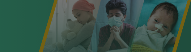
Craniosynostosis treatment often involves complex surgeries, medical consultations, and post-operative care. In India, where healthcare costs can be significant, families facing the diagnosis & treatment of Craniosynostosis often find themselves burdened with high medical expenses. The financial strain on families can be overwhelming. Crowdfunding provides a platform for individuals, friends, and communities to come together, pooling resources to help cover these substantial costs. It’s not just about financial assistance; it’s a collective effort to show care and solidarity.
Some families may be in remote areas with limited access to specialized medical facilities. Crowdfunding allows them to reach a global audience, increasing the chances of raising enough funds for the child’s treatment, regardless of geographical constraints.