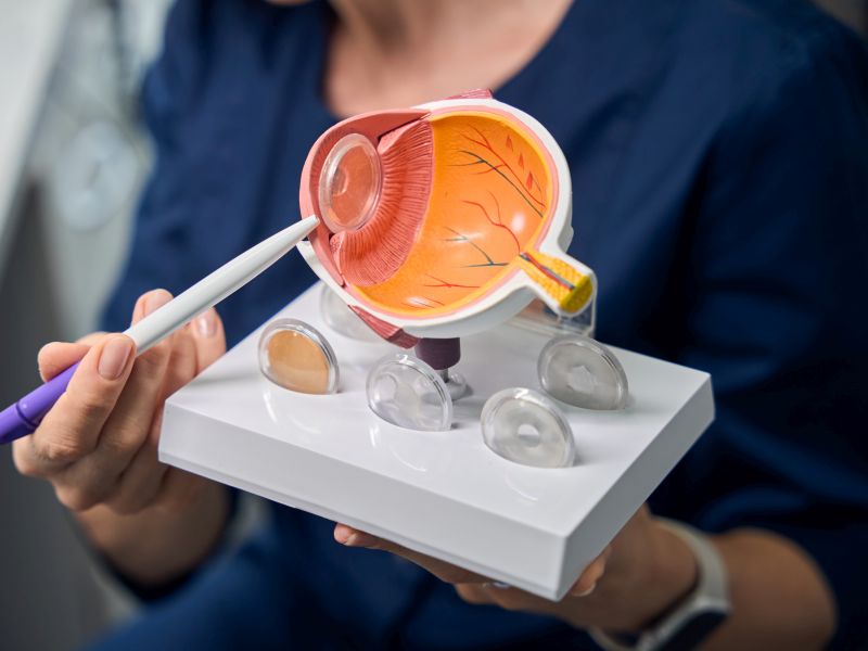
Retinal detachment is a severe eye condition that can cause vision loss if not treated instantly. It happens when the retina, the thin coating of light-sensitive tissue at the back of the eye, gets separated from its normal position. Since the retina plays a crucial role in processing visual information & sending it to the brain, any disruption can severely affect your ability to see.
When retinal detachment occurs, it’s crucial to seek medical help right away. Surgery is often the best way to repair the retina and prevent permanent vision damage. There are different types of surgical procedures available, depending on the severity and type of detachment. In this blog, we’ll explore retinal detachment, the importance of timely treatment, and the surgical options that can help save your vision.
Table of Contents
Retinal Detachment Causes

It is a serious eye condition that develops when the retina, the slim cover of tissue at the back of the eye, separates from the layer underneath it. This separation can lead to vision problems if not treated quickly. There are 3 main kinds of retinal detachment, each caused by different factors:
1. Rhegmatogenous Retinal Detachment
This is the most common kind of retinal detachment, and it happens due to a tear or hole in the retina. Here’s how it works:
– Vitreous gel shrinkage: Inside the eye, there’s a clear, gel-like substance called the vitreous. As people age, this gel can shrink or become more liquid, pulling away from the retina. In some cases, this can create a small tear or hole.
– Fluid buildup: Once there is a tear, the fluid from the vitreous gel can seep through the hole and gather behind the retina. This fluid buildup causes the retina to separate from the back of the eye, leading to detachment.
Risk factors:
– Aging (usually after 50)
– Severe nearsightedness (myopia)
– Eye injuries
– Previous eye surgeries, like cataract removal
– Family history of retinal detachment
Causes:
– Aging: As we age, the gel-like fluid inside the eye (called the vitreous) shrinks and can pull on the retina, causing a tear.
– Eye injuries: Trauma or accidents that affect the eye can sometimes lead to a tear in the retina.
– Severe nearsightedness: People with very poor vision are at higher risk because their eyes are often more elongated, which puts stress on the retina.
2. Tractional Retinal Detachment
This kind of retinal detachment happens when scar tissue on the retina’s surface contracts, pulling the retina away from its normal position. It is less common than rhegmatogenous detachment and is usually linked to medical conditions, especially those that influence the blood vessels in the retina.
Common causes:
– Diabetic retinopathy: In people with diabetes, a high blood sugar level can influence the blood vessels in the retina, leading to scar tissue formation, which may pull on the retina.
– Inflammation or infections: Certain eye diseases can cause scar tissue buildup.
3. Exudative Retinal Detachment
In this type, there is no tear or hole in the retina. Instead, fluid builds up under the retina due to other issues, pushing it away from the back of the eye.
Causes:
– Inflammation: Swelling inside the eye due to conditions like uveitis can lead to fluid accumulation under the retina.
– Tumors: Some tumors in the eye can leak fluid, causing the retina to detach.
– Leaking blood vessels: Conditions such as central serous retinopathy, where blood vessels leak fluid behind the retina, can also lead to detachment.
Other Factors That Can Contribute to Retinal Detachment:
– Eye injury or trauma: A hard blow or injury to the eye can sometimes cause the retina to detach, especially if there’s a tear.
– Eye conditions: Disorders like lattice degeneration (retina thinning) can make detachment more likely.
– Previous retinal detachment: If you’ve had a detachment in one eye, your risk increases in the other eye.
Each type of retinal detachment can lead to serious vision problems, so getting medical help immediately is important if you notice symptoms like sudden flashes of light, floaters (small spots in your vision), or a shadow across part of your visual field. Early detection & treatment are crucial to preventing permanent vision loss.
Understanding the causes of retinal detachment is vital because early detection & treatment can prevent severe vision loss. If you experience symptoms like sudden flashes of light, floaters (tiny dark spots or shapes in your vision), or a shadow covering part of your sight, it’s essential to seek medical attention immediately to check for retinal detachment.
Retinal Detachment Treatment
Several retinal detachment treatments are available, each with its own method and cost implications. Here are the most common treatments:
1. Pneumatic Retinopexy: This is a less invasive process where a gas bubble is injected into the eye. The bubble pushes the retina back into position. The patient must preserve a specific head position for a few days to keep the bubble in contact with the retina. This procedure is generally less expensive, depending on the hospital and city.
2. Scleral Buckle: This involves placing a flexible band around the eye, which indents the wall of the eye & relieves some of the force induced by the vitreous pulling on the retina. The scleral buckle is left in place permanently. The cost of scleral buckle surgery can vary widely but is typically more expensive than pneumatic retinopexy.
3. Vitrectomy: In this surgery, the vitreous gel pulling on the retina is removed & replaced with a gas bubble or oil to flatten the retina. Eventually, the body’s fluids will replace the gas or oil. Due to its complexity, a vitrectomy is usually more costly than the other procedures.
The price for these treatments can vary based on the hospital, the city, and the specific circumstances of the patient’s condition. In India, the price for retinal detachment treatment can range from approximately INR 30,000 to INR 1,50,000. For instance, the cost of retinal detachment treatment using laser surgery or cryopexy is between INR 3,000 and INR 5,000 per eye. The cost of retinal detachment treatment using other surgical methods in Mumbai is between INR 50,000 and INR 70,000. In Delhi, the price ranges from INR 45,000 to INR 65,000.
Symptoms To Watch Out For Indicating Potential Retinal Detachment
1. Sudden Appearance of Floaters
Floaters are small dark spots, shapes, or squiggly lines that drift across your field of vision. They are often described as tiny specks, cobwebs, or dust-like particles that move when you shift your eyes. While occasional floaters are normal and usually harmless, a sudden increase in the number of floaters can signify retinal detachment. This happens because the vitreous gel inside the eye may pull on the retina, causing a tear, which leads to more floaters appearing.
2. Flashes of Light (Photopsia)
Seeing flashes of light, especially in your peripheral vision, can be another symptom of retinal detachment. These flashes may appear like lightning streaks or bright sparks and are more noticeable in dim lighting or when your eyes are closed. The flashes occur because when the retina tears or detaches, it can send abnormal signals to the brain, creating the illusion of light.
3. A Shadow or Curtain Over Part of Your Vision
One of the more serious signs of retinal detachment is a dark shadow or curtain-like shape appearing in your vision. This shadow typically starts from the side (peripheral vision) and may gradually move toward the center of your sight. It might feel as if part of your field of vision is being blocked or covered. This symptom indicates that the retina is detaching and needs immediate medical attention.
4. Blurry or Distorted Vision
It could be an alarming sign of retinal detachment if your vision suddenly becomes blurry or wavy. This distortion may make straight lines look curved or bent, and your overall vision may feel as if it’s being affected by a “film” or cloudiness. As the retina separates, the quality of your vision declines because the retina is no longer functioning properly to transmit clear images to the brain.
5. Loss of Peripheral Vision
Peripheral vision is the ability to see things not directly in front of you. If you notice a gradual loss of peripheral vision—like tunnel vision where your central vision is intact but your side vision is disappearing—it could be due to retinal detachment. This happens because the outer edges of the retina are often the first to detach.
6. A Sudden Decrease in Vision
In some cases, people may experience a sudden, sharp decrease in their overall ability to see clearly. This can range from partial vision loss to complete blindness in one eye. A sudden change like this should never be ignored, as it could signal a serious problem, such as a large portion of the retina detaching.
When to Seek Medical Help:
If you experience any of these symptoms, it’s important to act quickly. Retinal detachment can progress rapidly; the longer it goes untreated, the higher the possibility of permanent vision loss. Eye doctors can use special tools to examine your retina and confirm whether it’s detached or at risk of detaching. Early treatment, often involving surgery, can help reattach the retina and prevent further damage.
Understanding Retinal Detachment Surgery
Retinal detachment surgery is a critical process performed to address the separation of the retina from its underlying support tissue in the eye. If left untreated, this medical condition can lead to permanent vision loss. The surgery aims to reattach the retina, restoring as much vision as possible and preventing further deterioration.
Pre-Surgery Steps
Before undergoing retinal detachment surgery, patients are typically advised to:
– Undergo a thorough eye examination.
– Cease taking certain medications that could affect the surgery’s outcome.
– Arrange for someone to accompany them to the hospital, as driving post-surgery will not be possible.
– Fast for a specific period before the surgery, usually from midnight on the day of the procedure.
Pre-Surgical Consultation
During the pre-surgical consultation, patients can expect:
– A detailed discussion about the surgical procedure, including the risks and benefits.
– Instructions on pre-operative preparations.
– An explanation of the anesthesia process and what will happen during the surgery.
The Surgery Experience
On the day of the surgery, patients should be prepared for:
– The administration of anesthesia, which could be local or general, depending on the case and patient comfort.
– The duration of the surgery can differ but often lasts between one and two hours.
– Post-operative care instructions that will be crucial for recovery.
Post-Surgery Recovery
After the surgery, the recovery process involves:
– Wearing an eye patch or shield to cover the eye.
– Using prescribed eye drops to prevent infection & control inflammation.
– Adhering to specific activity restrictions, such as avoiding strenuous exercises or heavy lifting.
– A recovery timeline can range from a few weeks to several months, depending on individual cases and the type of surgery performed.
Managing Potential Complications
Complications are rare but can include:
– Infection or bleeding within the eye.
– Increased intraocular pressure.
– Recurrence of retinal detachment.
– Cataract formation in some cases.
Patients are instructed to follow their surgeon’s instructions closely and attend all follow-up appointments to manage these risks effectively.
Long-Term Outcomes and Vision Restoration
The success rates for retinal detachment surgery are high, with over 90% of surgeries resulting in successful reattachment of the retina. However, the final visual outcome may vary based on the extent of the detachment and the time elapsed before surgery.
Rehabilitation and Visual Aids
Post-surgery, some patients may require visual aids or rehabilitation services to maximize their visual function. This can include:
– Special glasses or magnifiers.
– Adaptations in the living environment to enhance safety and independence.
– Support from low vision specialists to learn new strategies for managing reduced vision.
Conclusion
Retinal detachment is a severe eye condition that needs immediate medical attention to prevent permanent vision loss. With timely diagnosis and surgery, the retina can often be repaired, restoring vision. However, the cost of retinal detachment surgery in India can be high, especially for those without adequate insurance or savings.
This is where a fundraising platform can make a difference. By creating a campaign, individuals can seek support from friends, family, and even the wider community to help cover surgery costs. These platforms provide an accessible way to raise the necessary funds, ensuring that financial barriers do not prevent someone from receiving the urgent medical care they need to protect their vision.


